When you visit the dentist or orthodontist for the first time in a while, one of the first steps is getting an x-ray. We think of it as normal today, but this process wasn’t widely used for dental purposes until as recently as the 1950s!
Dr. Waters and Dr. Clayton at Innovative Orthodontics can tell you how these x-rays work (there’s more to it than you think) and help you answer the question, “Why do orthodontists use x-rays?”
The Era Before X-Rays
You might wonder what orthodontists used prior to having this technology. Well, orthodontists could take exterior photographs to document and track a patient’s condition during treatment but had limited access to internal information. The closest they could get was through the use of panoramic radiographs, which are wide-format images that are significantly less detailed than our modern x-rays.
Other methods included bite analysis and taking molds and impressions of the mouth. They would also take plaster models at regular intervals to measure progress in the teeth and jaw during orthodontic treatment.
In other words, it wasn’t as easy then as it is today.
Explaining The Process
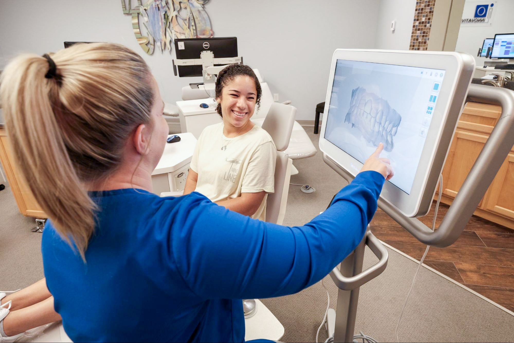
Radiographs are another term for dental x-rays. Electromagnetic radiation is used for this now common diagnostic tool, allowing us to take pictures of bones, teeth, and surrounding oral and facial structures. Curious how it all works? We can break it down for you:
- Our X-ray machine produces controlled bursts of radiation. One of our excellent dental assistants will usually operate this device before one of our orthodontists enters.
- Before snapping any images, we’ll drape you in something called a lead apron. This effectively blocks your vital organs from ionizing radiation, covering your chest and torso. We should note that modern X-ray technology limits a patient’s exposure to radiation better than it used to.
- We’ll place a digital sensor in your mouth and capture images. It can be positioned to take photos of the whole mouth or just to focus on areas of concern. Film was used for this purpose in the past and would need to be developed in a lab for viewing.
- Once the machine is activated, a focused beam of exposure is directed toward the sensor. Softer tissues like gums and cheeks allow more x-rays to pass through, creating darker areas on the image, while bones and teeth absorb more of the x-rays due to being dense, resulting in lighter areas on the final image.
- When the image appears on-screen, Dr. Waters or Dr. Clayton will examine it closely to determine how to proceed with treatment planning, or how to adjust an existing plan if needed.
- Extraoral x-rays capture the holistic oral and facial structure and are used to assess the head and jawbone.
- Intraoral x-rays capture the interior of the mouth and make small, hidden areas visible.
Using X-Rays As Orthodontists
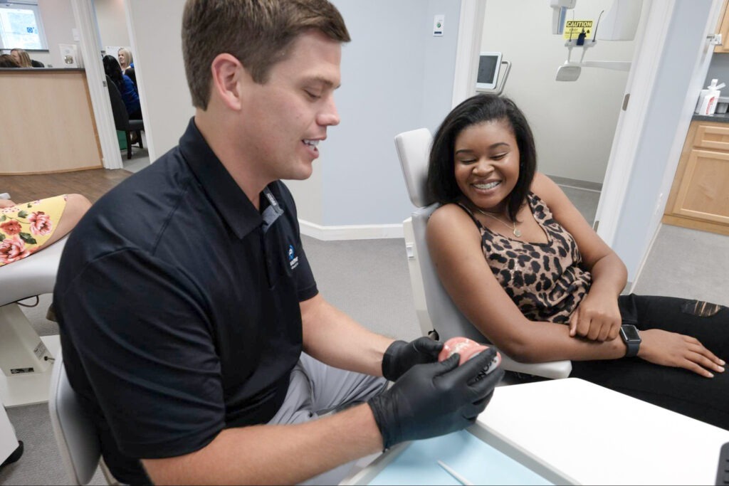
Now that you know the way it all works, we can talk about why and how our expert team uses x-rays in our three Georgia offices.
-
- Early Diagnosis of Orthodontic Issues: Pinpointing orthodontic problems in their earliest onset stages—hopefully before they become visibly apparent—is always preferable. Treatments are best when they’re interceptive and can prevent issues from getting more tedious.
- Assessment of Bite Problems: Underbites, overbites, crossbites, and open bites (otherwise known as malocclusion) are conditions we frequently see as orthodontists. X-rays show us the upper and lower jaws and the alignment of teeth so that we can effectively choose the best treatment options.
- Assessment of Tooth and Jaw Position: Probably the most notable part of this technology is that it provides us with detailed images of your teeth, jawbone, and the surrounding structures, which aren’t plainly visible to the eye. These images allow us to see the position, growth, and alignment of teeth and jaws in our patients across Georgia.
- Identification of Impacted Teeth: Impacted teeth are either blocked from eruption or simply don’t erupt correctly. Watching out for impacted teeth is important for our planning treatments, especially in cases where oral surgery might be required.
- Bone Health: When our treatment plan involves the repositioning of teeth, bones must be capable of supporting these adjustments. We can use x-rays to analyze the density of the bones involved in any procedure.
- Evaluation of Dental Development: X-rays help us keep records showing the development of teeth, most notably in children and teens, sometimes showing progress over a number of years. This also helps our team observe any problems with permanent teeth that are coming in.
- Treatment Verification: Post-treatment x-rays, let us confirm that your teeth and jaws have been realigned according to plan!
We’re X-tra Excited to Meet You!
So, long story short—our job at Innovative Orthodontics relies heavily on x-rays! We use all sorts of technology in our treatment processes and are happy to help answer any questions you might have. Whether you’re in Savannah, Rincon, or Pooler, you can schedule your free consultation with us today or give us a call at (912) 800-0294!